Wednesday, July 25, 2007
Monday, July 16, 2007
Unit 3 Ethical Issue- Exercise
Now a days it is being made easier for society to obtain things without working hard for them. Drive throughs with menus high in sugar and fat and down time filled with sitting and minimal activity. Obesity has become a side affect of a society that want's everything the quickest way possible while exerting the least amount of effort. Today there are tons of diet pills and surgeries on the market that are advertised in a way to seem like a quick fix. In reality, exercise takes time and dedication, but the benefits can be long lasting and life lengthening. Also in todays society, it seems that people are working alot more and when they have time off they want to spend it relaxing. Fewer people are spending time outdoors and more time with sedentary entertainment.
No matter the reasons everyone has the ability to make personal choices. Some countries are trying to encourage physical activity and healthier lifestyles which is a change in positive direction, but people are going to have to make the decision to participate in these changes. It is much easier to put exercise off or make excuses, but even light activity every day (taking the stairs instead of the elevator) can have a positive effect and become part of daily routine. Personally I have to make a conscious effort to make time to work out weekly. Between working 72 hours a week and other commitments every week there are many times the last thing I want to do is go to the gym; so I mix it up. In the mornings when I get off work, before I can sit down and fall asleep I take my three great danes for a 2 mile walk. This is minimal work, but none the less it is still physical activity and I usually feel better when I am done. Pretty soon this type of thing is just habit and not something that has to be forced
In conclusion, there a numerous benefits to exercises and also numerous excuses why people don't do it. In the end, I feel that you can not just blame society, but the individuals in society,it comes down to personal motivation and people are going to have to take the first steps themselves to exercise, eat well and overall lead healthier lives.
Works Cited
Mader, Sylvia. Human Biology 10th ed
Modifying the Environment to Reduce Obesity www.ehponline.org/docs/2005/7812/7812.html
Unit Three Lab- Limb
The following is a list of materials used and what they represent
- Hedger handles- Humerus, radius and ulna

- Popcicle sticks- elbow joint (trochlea),myosin
- Red Yarn- biceps brachii
- Pipe cleaners- cross bridges of myosin (blue), Actin (white), Axons (white), dendrite (white), T tubules (green)
- Styrofoam balls- different neurons
- Electrical tape- myelin sheath
- Hot glue- sensory receptors
- Wire- Z lines
- Clear tubing- sarcolemma, myofibril (with red lines)
- Markers and paint- used to add color to myofibril and axon terminals


This first picture is of the bone structure showing how the humerus connects to the radius an ulna by the elbo joint. This is a hinge joint and can only bend one way. The joint is represented by a roll of popsicle sticks. They represent the trochlea and capitulum .
The second picture shows the red yarn representing the biceps  brachii. It is attatched to the top of the humerus and scapula (origin) and also to the radius (insertion).
The third and fourth picture show the sarcomeres in a myofibril when they are relaxed and also when they are contracted. The sarcomeres contain actin and m yosin. When the im
 pulse travels down the t tubules (fifth picture), calcium is released into the sarcoplasmic reticulum causing the sarcomeres to contract (actin slides past myosin) and shorten.
 d myofibril with z lines.
The last picture is of the different types of neurons, their axons, dendrites, and m
 yelin sheath. The neurons are responsible for taking impulses from the CNS, summing up these impulses, distributing them to and effector to carry out the response.
In conclusion this model shows the different units involved in muscle movement from the neurons all the way up to bones and muscles. This model was a good learning tool because when I actually had to make these aspects I had to think about their functions and understand how they worked individually and then how they all tied together.
Unit 3, Topic 2 Online Lab- Muscles
This lab will show how temperature and muscle fatigue affect our muscles ability to contract. We will see first hand the differences in the rate they are able to contract when exposed to cold temperatures and after fatigue. These differences occur due to vasoconstriction and muscle fuel. Muscles use ATP as an energy source. ATP is created either anaerobically (only a limited amount) or aerobically (using oxygen) carried in the blood stream. Aerobic ATP lasts longer, but can only be produced so fast.
1. When I clenched my teeth, my cheek muscle became rigid as opposed to soft when my jaw is relaxed
2. The length of the bicep shortened when the arm is bent. This is due to the muscle contracting and pulling on the lower arm to move it. The origin is where the tendon connects to the top of the humerus and the insertion is where the tendon connects to the ulna (this is the moveable bone) The biceps brachii contract while the triceps brachii relax.
3. The circumference of the bicep enlarges when the fist is clenched tightly as opposed to a relaxed state
Temperature Number of Fists
Normal 29
After Ice Water 20
Trial # # of Sqeezes
1 40
2 40
3 39
4 35
5 35
6 35
7 33
8 30
9 29
10 28
2. The cold temperature had the effect of stiffening the hand muscles and making them slower to contract. This is due to the fact that with decrease in temperature, the smooth muscle in the blood vessels constrict. If blood vessels are constricted, less blood is circulating which means less oxygen. Oxygen is needed for the production of ATP which is the fuel used by cells and is also required for the sliding of the actin myosin filaments that causes muscle contraction.
3. Fatigue had the effect of being able to complete less contractions and with a weaker force in the same time period. This could be caused by the muscle cell not being able to receive enough calcium to induce the sliding of actin and myosin (contraction), due to rapidly contracting the hand with no rest in between. Also, with no rest, the muscles have no time to recover, or relax which could cause them to tire.
http://www.rogers.k12.ar.us/users/ehutches/musaction6.jpg
Works Cited
Mader, Sylvia. Human Biology 10th ed
About.com http://sportsmedicine.about.com/od/anatomyandphysiology/a/musclefatigue.htm
Sunday, July 15, 2007
Unit 3 Topic Two
- Overview of Skeletal System
- Bone Growth, Remodeling and Repair
- Bones of the Axial Skeleton
- Bones of the Appendicular Skeleton
- Articulations
Muscular System
- Overview of the Muscular System
- Skeletal Muscle Fiber Contraction
- Whole Muscle Contraction
- Muscular Disorders
- Homeostasis
 re five functions of the skeletal system
- Supporting the body
- Protecting soft body parts
- Producing blood cells
- Storing minerals and fat
- Permit flexible body movement
A long bone is made up of a shaft (has medullary cavity with walls made of compact bone and
Compact bone is made of osteons which are tubular units. In the osteon, osteocytes lie in lacunae. Osteocytes exchange nutrients and wastes with the blood vessels in central canal.
Spongy bone is made up of many thin plates (trabeculae) separated by unequal spaces and designed for strength. Often contain red bone marrow.
Cartilage is not as strong as bone, but more flexible. This is due to the matrix being gel-like with collagen and elastic fibers. These cells are called chondrocytes. Cartilage has no nerves and no blood vessels. There are three types of cartilage
- Hyaline- firm and flexible
- Fibrocartilage- stronger and used in support
- Elastic- most flexible (ear flaps and epiglottis)
Fibrous connective tissue is made of rows of fibroblasts that are separated by bundles of collagen fibers. This is the tissue that ligaments and tendons are made of. Ligaments connect bone to bone and tendons connect muscles to bone at joints.
Bone Growth, Remodeling, and Repair
The skeleton starts to develop when the embryo is only 6 weeks old. Bones are made of living tissue and are able to grow throughout a lifetime by responding to stress (change shape, size and strength)
- Osteoblasts- are bone forming, encourage the deposition of calcium salts into the matrix
- Osteocytes- are mature bone cells that maintain structure
- Osteoclasts- are bone absorbing cells that break down bone and deposit calcium and phosphate into the blood.
Ossification is formation of bone. There are two different types of ossification
- Intramembranous- bones develop between sheets of fibrous connective tissue. These cells become osteoblasts and are found in ossification centers. They secrete the organic matrix and when calcium salts are added this results in calcification. Trabecule of spongy bone are formed and a periosteum develops around the outside. Compact bone is created on the outside. Example include skull bones and flat bones
- Endochondral- This is when cartilaginous models are replaced by real bones. Bone formation occurs from the center of the model to the end
- Cartilage model- chondrocytes put down hyaline cartilage which is the model
- Bone collar- Osteoblasts from the new periosteum secrete the matrix and then it undergoes calcification creating the bone collar to cover the diaphysis
- Primary ossification center- blood vessels carry osteoblasts to the interior to lay down spongy bone
- Medullary cavity and secondary ossification sites- spongy bones absorbed by osteoclasts and creates the medullary cavity
- Epiphyseal plate- band of cartilage that remains between primary ossification centers and each secondary center. Allows limbs to increase in length. There are four layers. First is the resting layer which remains cartilage, then the proliferating layer where chondrocytes produce new cartilage. Third is the degenerating zone where cartilage cells die and last is the ossification zone where bone is formed. When these plates close, the bone can no longer grow in length
Hormones are chemical messengers produced by one part of the body, but acting on another part. Vitamin D is converted into a hormone for the intestinal tract to help it absorb calcium. Growth hormone stimulates the epiphyseal plate and general bone growth. Thyroid hormone promotes metabolic activity in the cells.
Bone remodeling (bone renewal) keeps bones strong, while bone recycling allows regulation of calcium in the blood. The parathyroid hormone accelerates bone recycling and calcitonin is the hormone that acts the opposite way of parathyroid.
Bone repair is needed if a bone is fractured or broken. This repair can take several months and works in the following steps
- Hematoma- blood from the ruptured vessels forms a mass of clotted blood in between the broken bones
- Fibrocartilaginous callus- tissue repair starts and this callus fills in the space bewtween the broken part for up to three weeks
- Bony callus- Osteroblasts produce trabeculae and convert the fibrocartilaginous callus to a bony callus the joins the bones together for 3-4 months
- Remodeling- osteoblasts build new compact bone and osteoclasts absorb spongy bone which creates a new medullary cavity.
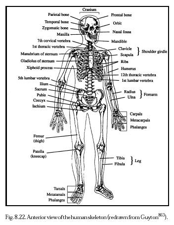
Bones of the Axial Skeleton
All of the skeletal bones are classified depending if they occur in the axial skeleton or appendicular skeleton. The axial skeleton is made of
Skull-
- Cranium ( protects the brain).
- Frontal bone (forms forehead),
- Parietal bone (sides)
- Occipital bone (base of the skull).
- Foramen magnum is the opening where the spinal cord passes and turns into the brain stem.
- Temperal bone (leads to middle ear),
- Sphenoid bone (extends across floor of cranium. all bones articulate with this one),
- Ethmoid bone (forms the nasal septum and orbits)
- Facial bones (mandible- only moveable bone, maxillae, zygomatic, nasal bones)
Hyoid Bone- only bone in the body that does not articulate with another bone. Attatched to temporal bones by muscle and ligaments and a membrane connects it to the larynx. Hyoid anchors the tongue and is place of attatchment for muscles used in swallowing
Vertebral Column- has 33 vertebrae that are named for their location in the vertebral column. First is the atlas (holds up the head). Second is the axis (rotation) then 12 thoracic, 5 lumbar, 5 sacrum, and 3-5 fused coccyx.
Rib Cage- also known as the thoracic cage is made of thoracic vertebrae, ribs, sternum, and cartilage. It protects the heart and moves with inspiration and expirtation.
Bones of the Appendicular Skeleton
These are bones in the pectoral and pelvic girdles and attatched limbs.
Pectoral girdle- there a right and left. Each one has a scapula, clavicle, glenoid cavity, rotator cuff, humerus, radius and ulna, carpal, metacarpals and phalanges.
Pelvic girdle- has two hip bones, pelvis, coxal bone ( ilium, ischium, pubis), pubic symphysis, femur, tibia, patella, fibula, tarsal, metatarsal and phalanges.
Articulations
Bones are joined together at joints (fibrous, cartilaginous, synovial) Most fibro
Muscular System
Overview of Muscular System
All muscles have the ability to contract causing movement. There are three ty
- Smooth muscle- spindle shaped cells with a single nucleus. Arranged in parrallel lines which form sheets. No striations. Located in walls of hollow internal organ and contraction is involuntary
- Cardiac muscle- form the heart wall. No nucleus, striated, tubular and branched which allows fibers to interlock. These fibers relax completely in between contractions which prevents fatigue. Involuntary contractions
- Skeletal muscle- fibers are tubular, have many nucleus' and are striated. They make up skeletal muscles and are voluntary.
Functions of the Skeletal muscles include
- Supporting the body- contraction opposes gravity and we remain upright
- Making bones move-muscle contraction results in movement
- Maintaining constant body temperature-contraction causes the breakdown of ATP which releases heat
- Assists movement in cardiovascular and lymphatic vessels- pressure of contraction keeps blood moving
- Protect internal organs and stabilize joints- pad bones, hold them together at joints and protect internal organs

Skeletal muscles are attatched to the skeleton. A muscle contains bundles of skeletal muscle fibers (fascicles). Each fiber is surrounded in connective tissue. Muscles are covered in fascia that goes past the muscle and becomes the tendon. These anchor a muscle to a bone. Usually each muscle works on the movement of one bone. These muscles work in pairs. Origin is on the stationary bone and insertion is on the bone that moves. Muscles can only pull, not push and this is why they work in pairs. A prime mover does most of the work with assistance from synergists and the antagonist is the muscle that works opposite the prime mover. One must relax while the oher is working.
The following terms help to characerize muscles
- Size- maximus, minimus, vastus, longus, brevis
- Shape- deltoid, trapezius, latissimus, terres
- Location- external, internal, frontalis,pectoralis, gluteaus,brachii, sub
- Direction of muscle fibers-rectus, orbicularis,transverse, oblique
- Attachment- sternocleidomastoid, brachioradialis
- Number of attachments-
- Action- extensor, adductor, flexor, masseter, levator
Skeletal Muscle Fiber Contraction
A muscle fiber is made up of a sarcolema(plasma membrane), sarcoplasm (cyt
Myofibrils run the length of the muscle fiber. Sarcomeres are units of myofibrils and extends betweeen the Z lines. Contains myosin and actin. The I band has only actin and the 
Thick filaments is hundreds of myosin molecules. Myosin is shaped like a golf club (the end is a cross bridge) Thin filaments is two intertwining strands fo actin. Sliding filaments let actin slide over myosin during contraction when the impulses travel down the T tubule into the sarcoplasmic reticulum where calcium is released. When sliding occurs, sarcomeres shorten and the myosin filaments break down ATP.
Muscle fibers contract when stimulated by motor neurons with there axons i
Whole Muscle Contraction
A nerve fiber with all the muscle fibers in innervates is a motor unit. This uni
Muscles use different fuel sources for energy and different ways to produce ATP during contraction. Glycogen and fat are stored in muscle while blood glucose and plasma fatty acids come from the blood. Muscles acquire ATP by the creatine phosphate pathway, fermentation, can cellular respiration
Fast twitch fibers metabolize anaerobically and are designed for strength due to the fact their motor units contain many fibers and provide explosions of energy. They develop maximum tension more rapidly, but are more susceptible to accumulation of lactate
Slow twitch fibers have more endurance and produce energy aerobically causing them to tire when their fuel supply is gone. Contain myoglobin and lots of mitochondria. Surounded by capillary beds and draw more blood and oxygen.
Muscular Disorders
Common disorders
- Spasms- sudden, involuntary muscular contraction
- Cramps- strong, painful spasms
- Strain- stretching or tearing a muscle
- Sprain- twisting of a joint, includes damage to ligaments, tendons, blood vessels, nerves
- Tendinitis- gliding motion of tendon impaired
- Bursitis- inflamation of bursa
Serious conditions
- Myalgia-achy muscles due to overuse or over stretching
- Muscular dystrophy- group of disorders characterized by progressive degeneration
- Myasthenia gravis- autoimmune disease characterized by weakness. Muscle contraction is impaired when the immune system accidentally produces antibodies that destroy ACh
- Amyotrophis lateral sclereosis- gradual loss of ability to walk, talk, chew, and swallow
Homeostasis
Muscle movement allows us to respond to environmental changes, allow us to eat, to digest that which we eat to provide cells with nutrients, breathe, circulate blood and lymph. Also the skeletal and muscle systems protect internal organs allowing them to carry out their part of homeostasis. The skelaton is largely involved in calcium homeostasis. Also blood cells are produced in bones in the red bone marrow where white blood cells are also produced. Muscles help maintain body temperature (shivering, goosebumps)
Works Cited
Mader, Sylvia. Human Biology 10th ed
Frolich Powerpoint
Links for Pictures
1. http://www.web-books.com/eLibrary/Medicine/Physiology/Skeletal/long_bone.jpg
2. http://www.nanomedicine.com/NMI/Figures/8.22.jpg
3. http://www.pinkmonkey.com/studyguides/subjects/biology-edited/chap20/fig20_2.gif
4. http://picinfor.googlepages.com/02620Types20of20joints20found20in20the20human20body.jpg/02620Types20of20joints20found20in20the20human20body-full.jpg
5. http://virtualastronaut.jsc.nasa.gov/textonly/act21/images/musclemap.png
6. http://www.agen.ufl.edu/~chyn/age2062/lect/lect_19/146.gif
7. http://www.mhhe.com/biosci/esp/2001_gbio/folder_structure/an/m5/s5/assets/images/anm5s5_1.jpg
8. http://www.rogers.k12.ar.us/users/ehutches/musaction6.jpg
9. http://fig.cox.miami.edu/~cmallery/150/neuro/neuromuscular-sml.jpg
10. http://www.edcenter.sdsu.edu/cso/paper/image005.jpg
Saturday, July 14, 2007
Online Lab Topic One- Neurons
1. What is the electrode measuring?
The electrode is measuring the voltage of individual neurons and the
reactions when these neurons are stimulated (second picture shows
a t cell and the different reading obtained when the skin was
stimulated with the three different tools).
 2. Why use leeches in neurophysiology experiments?
Leeches are used because of their large accessible neurons. Also their simple system is easier to understand while at the same time relating to our complex system
3. What is the difference between a sensory and a motor neuron?
A sensory neuron is a nerve cell that sends impulses to the central nervous system once a receptor has been stimulated. A motor neuron is a nerve cell that sends impulses away from the central nervous system and innervates effectors like muscles and glands

4. Do you think a leech experiences pain? What is pain?
Pain is considered to have two aspects, one being physical hurt, and the second being emotional suffering. Nociceptive nerve detect injury causing stimuli have been identified in the leech, but is thought that the leech and other invertebrates are only capable of stimulus response reactions and do not have the brain system to process pain http://www.wellcome.ac.uk/en/pain/microsite/culture2.html
5. What were the two most interesting things about doing this lab?
The two most interesting things about this lab was the way you could actually
use the tools to dissect the leach, and also being able to watch the reactions when the
leach was stimulated with the different items.
6. Anything you found confusing or didn't like about the lab?
The lab was pretty straight forward, and easy to follow. The directions
were clearly stated and easy to understand, overall this was a fun lab
Thursday, July 12, 2007
Unit 3 Topic One Review
Nervous System
- Overview of the Nervous System
- The Central Nervous System
- The Limbic System and Higher Mental Functions
- The Peripheral Nervous System
- Drug Abuse
Senses
- Sensory Receptors and Sensations
- Proprioceptors and Cutaneous Receptors
- Senses of Taste and Smell
- Sense of Vision
- Sense of Hearing
- Sensce of Equilibrium
- Receive Sensory Input- sensory receptors respond to interal and external stimuli. Nerve impulses travel by the peripheral system to the central system.
- CNS Performs Integration- the central nervous system takes in all the information
- CNS Generates Motor Output- nerve impulse from the central system travel by the peripheral system to muscles and glands.
Nervous tissue is made up of neurons (transmit nerve impulses) and neuroglia (supports and nourishes the neurons). There are three types of neurons:
- Sensory Neuron-takes the impulses from a receptor (structure
 that detects changes)to the central system
 - Interneuron- lies completely in the central system. These receive input from each other and sensory neurons. They then take into account all of the impulses received and communicate with motor neurons
- Motor Neuron- takes the impulses from the central system to an effector ( this is a muscle fiber or gland that carries out a response to the environmental changes)

A neuron physically consists of a cell body, dendrites and an axon. The cell body has a nucleus and organelles. Dendrites are extensions used to receive signals from receptors and other neurons. An axon conducts nerve impulses. An axon in nerves is called a nerve fiber.
Most axons are covered in a myelin(lipid substance) sheath. This can be formed by neurolgia known as Schwann cells. These cells contain mylein in the plasma membranes. Schwann cells are in the peripheral system. In the central system oligodendrocytes perform the same function. The myelin sheath is developed when the oligodendrocytes wrap around an axon many times. Nodes of Ranvier are spaces where there is no myelin sheath. This occurs because each neuoglia cel can only cover a portion of an axon. Grey matter in the central system is due to a lack of myelinated axons, and white matter is due to myelinated axons. 
Nerve impulses relate information in the nervous system. These impulses can be recorded and studied by using a voltmeter. The resting potential is when the axon is not conducting any impulses. (inside of neuron is more negative then outside: Na+ greater on outside and K+ greater on inside due to sodium- potassium pump). Action potential is when polarity rapidly changes across the membrane. This occurs when there is an impulse. Na+ moves in and this is depolarization. Then K+ moves out and this is repolarization. Each action potential will create another one down the length of the axon.
Each axon branches into "fine endings tipped by a small swelling called an axon terminal" These terminals lie close to the dendrite or cell body of another neuron. This is called a synapse. A synaptic cleft is the space between the sending and receiving neurons. Transmission is c arried out by neurotransmitters that are stored in synaptic vesicles in axon terminals.
- Nerve impulses that are traveling along the axon reach the terminal.
- Calcium ions enter the terminal and stimulate synaptic vesicles and these merge with the sending membrane.
- Neurotransmitters are released into synaptic cleft where they diffuse across into the receiving membrane and then they bind with specific receptor proteins.
Some known neurotransmitters include acetylcholine, norepinephrine, dopamine, serotonin, glutamate, and gamma aminobutyric acid.
Central Nervous System
Sensory information is received and motor control initiated in the central nervous system. The spinal chord is surrounded by vertebrae while the brain is protected by the skull. Along w ith bone these structures are also protected in membranes called meninges. Spaces in between are filled with cerebrospinal fluid to cushion the central system. The central system has two types of nervous tissue: white (myelinated axons) and grey (cell bodies and non myelinated fibers).
The spinal cord runs from the base of the brain all the way down the vertebral canal. Spinal nerves project from the cord between the vertebrae, and intervertebral disks separate the vertebrae. Communication between the brain and peripheral nerves occurs through the spinal cord. The left side of the brain controls your right side of the body and vice versa. When the brain starts voluntary control of the limbs, motor impulses starting in the brain pass down the descending tracts to the spinal cord and then to our muscles through muscles fibers.
Reflex actions occur when a stimulus makes the sensory receptors generate a nerve impulse. This impulse travels to sensory axons to the spinal cord. Motor axons then cause skelatal muscles to contract. The spinal cord works the same way for internal organs.
Four ventricles make up the brain: two lateral ventricles, the third  ventricle and the fourth ventricle.
The cerebrum is the largest portion of the brain. This is the last stop for sensory input and can communicate with other parts of the brain. The cerebrum is divided into two hemispheres and sulci divide these into lobes (frontal, parietal, occipital, temporal). Cerebral cortex is an outer layer of grey matter covering the hemispheres (is responsible of sensation, voluntary movement, and thought processes)
The cerebellum is located under the occipital lobe and is separated by the fourth ventricle. It is broken into two sections joined by a median portion. Sensory input from the eyes, ears, joints,and muscles come here. It then performs integration and sends motor impulses throught the brain stem to skelatal muscles. (maintains posture and balance)
Brain stem contains mid brain, pns, and medulla oblongata. The midbrain is a relay station between cerbrum and spinal cord and cerbellum. Pons holds axon bundles and regulates breathing rate, and head movements. Medulla oblongata has reflex centers and regulates heartbeat, breathing, blood pressure. It is located superior to the spinal cord.
The Reticular Formation is a complex network of nuclei and receives sensory signals and sends them onward to higher centers. This is responsible for people being alert.
The Limbic System and Higher Mental Functions
The limbic system is a group of linked structures within the cerebrum and is involved in emotions and higher mental functions. It blends our primitive emotions with higher mental functions. Two important structures are the amygdala (causes experiences to have emotional overtones) and the hippocampus (involved in learning and memory)
Memory is the ability to hold a thought or recall events from the past. Learning is when we retain and use the past memories. There is short term memory (in the prefrontal area) and long term memory (mixture of sematic memory, and episodic memory) Long term memory is stored in pieces throughout the sensory association areas of the cerebral cortex. Language is dependent on semantic memory, so if there are disruptions of either memory or language pathways, it could contribute to an inability to comprehend the environment and effectively use speech.
The Peripheral Nervous System
This system is outside of the central nervous system and contains nerves. Cranial nerves arise from the brain and spinal nerves arise from the spinal cord. Nerves are composed of axons. There are 12 pairs of cranial nerves some are sensory and some are motor. The vagus nerve works with the medulla oblongata to control internal organs. Spinal nerves are in 31 pairs. The spinal nerve roots physically separate axons of sensory neurons from motor neurons. Spinal nerves are mixed nerves.
The somatic system is one division of the peripheral system. These nerves relate to the skin, skelatal muscles and tendons. Nerves take info from external sensory receptors the the central system and motor commands from the central system to the skelatal muscles. Reflexes also occur in the somatic system.
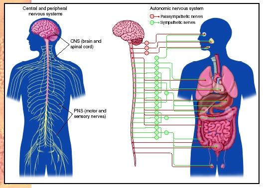
The autonomic system regulates smooth muscle and gland activity along with cardiac activity. The autonomic system is broken down into the sympathetic and parasympathetic systems. They both
- Automatically function in an involuntary manner
- Innervate internal organs
- Utilize two neurons and one ganglion for each impulse
These reflex actions (blood pressure and breathing rate) are essential to homeostasis. Sympathetic is used in fight or flight responses. It inhibits the digestive tract and releases norepinephrine. Parasympathetic promotes internal responses in a relaxed state. (digestion, pupil dialation). Known as the rest and digest system.
Drug Abuse
Drugs affect the nervouse system, therefore altering mood and or emotions. They affect the limbic system and either increase or decrease actions of particular neurotransmitters. Drug abuse is classified by someone taking a dose level and under circumstances that can increase potential of a harmful effect. Some of the more common drugs seen today include, alcohol, nicotine, cocaine, methamphetamine, heroin and marijuana.
Senses
Sensory Receptors and Sensations
Sensory receptors are specialized dendrites that detect specific stimuli. Exteroceptors detect from the outside of the body(environmental conditions) and interoceptors detect from the inside of the body. (these effect homeostasis and are controlled by the  negative feedback mechanism.)
There are four categories of sensory receptors
- Chemoreceptors- respond to chemical substances
- Pain receptors- these are a type of chemoreceptors and respond to the chemicals that damaged tissue releases
- Photoreceptors- respond to light energy
- Mechanoreceptors- these are stimulated by mechanical forces and can result in pressure.(sense of touch, hearing)
- Thermoreceptors- these are in the skin and hypothalamus and respond to changes in temperature.
Our sensory receptors respond to our external environment and send nerve impulses to the cerebral cortex where sensation (consious perception) occurs. All sensory receptors initiate nerve impulses that have been integrated (summing of the signals) When these reach the brain this is when sensation occurs. With out our sensory receptors, we would not receive info about our external and internal environments and homeostasis would not be possible.
Proprioceptors and Cutaneous Receptors
Muscle, joint, tendons and internal organ sensory receptors send their impulses to the spinal cord. After the spinal cord they go to the somatosensory areas. There are three types of general sensory receptors.
- Proprioceptors- are a type of mechanoreceptor that maintain muscle tone and the body's equillibrium and posture. They know degree of muscle relaxation, contraction, tension and movement.
- Cutaneous Receptors- located in the dermis of the skin, and effectivly making the skin sensitive to pressure, pain, and temperature. Fine touch gives information about the location, shape, size and texture of the touch (Meissner corpuscles, Merkel discs, and root hair plexus). Pacinian corpuscles and Ruffini endings are sensitive to pressure
- Pain Receptors- these are sensitive to chemicals released by damaged tissue. Referred pain is when the stimulation of internal pain receptors is felt as pain from the skin or the internal organs
Senses of Taste and Smell
Taste and smell are chemical senses because their receptors are sensitive to molecules in food and air. Chemoreceptors (plasma membrane receptors that bind to certain molecules) communicate by sensory nerve fibers with the respiratory center in the medulla oblongata.
Humans have around 3,00o taste buds on the tongue. The four primary types of taste are sweet sour, salty, and bitter. Molecules bind to receptor proteins on microvilli where nerve impulses are generated in the sensory nerve fibers and sent to the brain.
80-90% of what we think of as taste is actually due to smell. Smell is dependent on up to 20million olfactory cells in the roof of the nasal cavity. An odors signature in the olfactory bulb is determined by which neurons it stimulates. Olfactory cells decline and become less sensitive as we grow older.
Sense of Vision
The eyeball is made up of three layers:
- Sclera- white and fibrous. The cornea consists of transparent collagen fibers and is the window of the eye.
- Choroid- vascular and absorbs stray light rays. In the front it becomes the iris which regulates the size of the pupil (where light enters). Behind the iris is the ciliary body which controls the shape of the lens for near and far vision. The lens divides the eye into two compartments, the anterior compartment(filled with aqueous humor) and the posterior compartment.
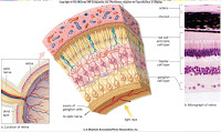
- Retina- located in posterior compartment (filled with vitreous humor). Contains rod cells and cone cells. Rods are sensitve to light but do not register color, and cones require bright light to recognize the different wavelengths (color). Fovea centralis is area of densely packed cells, and light is focused here when looking at an object. Sensory fibers from
 the retina form the optic nerve which in turn takes the impulses to the visual cortex.


The cornea is where focusing starts, and it continues through the lens and humors. Visual accomodation happens for close vision by the lens becoming more round to bring the object into focus.
Sense of Hearing
The two functions of the ear are hearing and balance. All of the sensory receptors of the ear contain hair cells with sereocilia sensitive to mechanical stimulation. These are mechanoreceptors. The ear is divided into three parts
- Outer- contains the pinna and auditory canal
- Middle- starts at the tympanic membrane and ends at the oval and round windows. The ossicles between the tympanic membrane and oval windows are: malleus, incus, stapes. The auditory tube allows the equalization of air pressure
- Inner- filled with fluid and broken down into semicircular canals, vestibule, and cochlea.
Hearing starts when sound waves enter the auditory canal and travel by vibrations to the tympanic membrane. The malleus takes this pressure and passes it using the incus to the stapes which hits the oval window membrane and passes the pressure to the fluid of the cochlea. In the cochlea the spiral organ has little hair cells and a tectorial membrane. Nerve impulses begin in the cochlear nerve and go to the brain. It is thought that the brain interprets tone based on the distribution of the stimulated hair cells
Sense of Equilibrium
The vestibular nerve works to achieve equilibrium and takes the nerve impulses to the cerebellum and brain stem. There are two types of mechanoreceptors involved in equilibrium
- Rotational- mechanoreceptors in semicircular canals detect angular and rotational movement. The three canals are arranged to each cover one dimension of space. The ampulla of each canal is enlarged and covered in little hair cells. As fluid in the canals moves over the ampulla, the hairs bend and the pattern of impulses being carried to the brain is changed.
- Gravitational- mechanoreceptors in the utricle and saccule detect verticle and horizontal movement. These are two membraneous sacs containing little hair cells in an otolithic membrane. When the head moves one way or another the otoliths are displaced and hair cells bent sending info to the brain. The brain uses the information it receives to maintain equilibrium by sending signals to the correct skelatal muscles to correct position
Works Cited
Mader, Sylvia. Human Biology 10th ed
Frolich powerpoint
Links to Pictures
1. http://www.virtualsciencefair.org/2004/visa4a0/public_html/brain6.gif
2. http://www.discoveryfund.org/images/Eye_Anatomy-Anat.jpg
3. http://www.biologyreference.com/images/biol_03_img0336.jpg