Nervous System
- Overview of the Nervous System
- The Central Nervous System
- The Limbic System and Higher Mental Functions
- The Peripheral Nervous System
- Drug Abuse
Senses
- Sensory Receptors and Sensations
- Proprioceptors and Cutaneous Receptors
- Senses of Taste and Smell
- Sense of Vision
- Sense of Hearing
- Sensce of Equilibrium
- Receive Sensory Input- sensory receptors respond to interal and external stimuli. Nerve impulses travel by the peripheral system to the central system.
- CNS Performs Integration- the central nervous system takes in all the information
- CNS Generates Motor Output- nerve impulse from the central system travel by the peripheral system to muscles and glands.
Nervous tissue is made up of neurons (transmit nerve impulses) and neuroglia (supports and nourishes the neurons). There are three types of neurons:
- Sensory Neuron-takes the impulses from a receptor (structure
 that detects changes)to the central system
 - Interneuron- lies completely in the central system. These receive input from each other and sensory neurons. They then take into account all of the impulses received and communicate with motor neurons
- Motor Neuron- takes the impulses from the central system to an effector ( this is a muscle fiber or gland that carries out a response to the environmental changes)

A neuron physically consists of a cell body, dendrites and an axon. The cell body has a nucleus and organelles. Dendrites are extensions used to receive signals from receptors and other neurons. An axon conducts nerve impulses. An axon in nerves is called a nerve fiber.
Most axons are covered in a myelin(lipid substance) sheath. This can be formed by neurolgia known as Schwann cells. These cells contain mylein in the plasma membranes. Schwann cells are in the peripheral system. In the central system oligodendrocytes perform the same function. The myelin sheath is developed when the oligodendrocytes wrap around an axon many times. Nodes of Ranvier are spaces where there is no myelin sheath. This occurs because each neuoglia cel can only cover a portion of an axon. Grey matter in the central system is due to a lack of myelinated axons, and white matter is due to myelinated axons. 
Nerve impulses relate information in the nervous system. These impulses can be recorded and studied by using a voltmeter. The resting potential is when the axon is not conducting any impulses. (inside of neuron is more negative then outside: Na+ greater on outside and K+ greater on inside due to sodium- potassium pump). Action potential is when polarity rapidly changes across the membrane. This occurs when there is an impulse. Na+ moves in and this is depolarization. Then K+ moves out and this is repolarization. Each action potential will create another one down the length of the axon.
Each axon branches into "fine endings tipped by a small swelling called an axon terminal" These terminals lie close to the dendrite or cell body of another neuron. This is called a synapse. A synaptic cleft is the space between the sending and receiving neurons. Transmission is c arried out by neurotransmitters that are stored in synaptic vesicles in axon terminals.
- Nerve impulses that are traveling along the axon reach the terminal.
- Calcium ions enter the terminal and stimulate synaptic vesicles and these merge with the sending membrane.
- Neurotransmitters are released into synaptic cleft where they diffuse across into the receiving membrane and then they bind with specific receptor proteins.
Some known neurotransmitters include acetylcholine, norepinephrine, dopamine, serotonin, glutamate, and gamma aminobutyric acid.
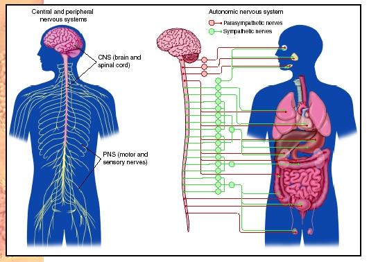
Central Nervous System
Sensory information is received and motor control initiated in the central nervous system. The spinal chord is surrounded by vertebrae while the brain is protected by the skull. Along w ith bone these structures are also protected in membranes called meninges. Spaces in between are filled with cerebrospinal fluid to cushion the central system. The central system has two types of nervous tissue: white (myelinated axons) and grey (cell bodies and non myelinated fibers).
The spinal cord runs from the base of the brain all the way down the vertebral canal. Spinal nerves project from the cord between the vertebrae, and intervertebral disks separate the vertebrae. Communication between the brain and peripheral nerves occurs through the spinal cord. The left side of the brain controls your right side of the body and vice versa. When the brain starts voluntary control of the limbs, motor impulses starting in the brain pass down the descending tracts to the spinal cord and then to our muscles through muscles fibers.
Reflex actions occur when a stimulus makes the sensory receptors generate a nerve impulse. This impulse travels to sensory axons to the spinal cord. Motor axons then cause skelatal muscles to contract. The spinal cord works the same way for internal organs.
Four ventricles make up the brain: two lateral ventricles, the third  ventricle and the fourth ventricle.
The cerebrum is the largest portion of the brain. This is the last stop for sensory input and can communicate with other parts of the brain. The cerebrum is divided into two hemispheres and sulci divide these into lobes (frontal, parietal, occipital, temporal). Cerebral cortex is an outer layer of grey matter covering the hemispheres (is responsible of sensation, voluntary movement, and thought processes)
The cerebellum is located under the occipital lobe and is separated by the fourth ventricle. It is broken into two sections joined by a median portion. Sensory input from the eyes, ears, joints,and muscles come here. It then performs integration and sends motor impulses throught the brain stem to skelatal muscles. (maintains posture and balance)
Brain stem contains mid brain, pns, and medulla oblongata. The midbrain is a relay station between cerbrum and spinal cord and cerbellum. Pons holds axon bundles and regulates breathing rate, and head movements. Medulla oblongata has reflex centers and regulates heartbeat, breathing, blood pressure. It is located superior to the spinal cord.
The Reticular Formation is a complex network of nuclei and receives sensory signals and sends them onward to higher centers. This is responsible for people being alert.
The Limbic System and Higher Mental Functions
The limbic system is a group of linked structures within the cerebrum and is involved in emotions and higher mental functions. It blends our primitive emotions with higher mental functions. Two important structures are the amygdala (causes experiences to have emotional overtones) and the hippocampus (involved in learning and memory)
Memory is the ability to hold a thought or recall events from the past. Learning is when we retain and use the past memories. There is short term memory (in the prefrontal area) and long term memory (mixture of sematic memory, and episodic memory) Long term memory is stored in pieces throughout the sensory association areas of the cerebral cortex. Language is dependent on semantic memory, so if there are disruptions of either memory or language pathways, it could contribute to an inability to comprehend the environment and effectively use speech.
The Peripheral Nervous System
This system is outside of the central nervous system and contains nerves. Cranial nerves arise from the brain and spinal nerves arise from the spinal cord. Nerves are composed of axons. There are 12 pairs of cranial nerves some are sensory and some are motor. The vagus nerve works with the medulla oblongata to control internal organs. Spinal nerves are in 31 pairs. The spinal nerve roots physically separate axons of sensory neurons from motor neurons. Spinal nerves are mixed nerves.
The somatic system is one division of the peripheral system. These nerves relate to the skin, skelatal muscles and tendons. Nerves take info from external sensory receptors the the central system and motor commands from the central system to the skelatal muscles. Reflexes also occur in the somatic system.
The autonomic system regulates smooth muscle and gland activity along with cardiac activity. The autonomic system is broken down into the sympathetic and parasympathetic systems. They both
- Automatically function in an involuntary manner
- Innervate internal organs
- Utilize two neurons and one ganglion for each impulse
These reflex actions (blood pressure and breathing rate) are essential to homeostasis. Sympathetic is used in fight or flight responses. It inhibits the digestive tract and releases norepinephrine. Parasympathetic promotes internal responses in a relaxed state. (digestion, pupil dialation). Known as the rest and digest system.
Drug Abuse
Drugs affect the nervouse system, therefore altering mood and or emotions. They affect the limbic system and either increase or decrease actions of particular neurotransmitters. Drug abuse is classified by someone taking a dose level and under circumstances that can increase potential of a harmful effect. Some of the more common drugs seen today include, alcohol, nicotine, cocaine, methamphetamine, heroin and marijuana.
Senses
Sensory Receptors and Sensations
Sensory receptors are specialized dendrites that detect specific stimuli. Exteroceptors detect from the outside of the body(environmental conditions) and interoceptors detect from the inside of the body. (these effect homeostasis and are controlled by the  negative feedback mechanism.)
There are four categories of sensory receptors
- Chemoreceptors- respond to chemical substances
- Pain receptors- these are a type of chemoreceptors and respond to the chemicals that damaged tissue releases
- Photoreceptors- respond to light energy
- Mechanoreceptors- these are stimulated by mechanical forces and can result in pressure.(sense of touch, hearing)
- Thermoreceptors- these are in the skin and hypothalamus and respond to changes in temperature.
Our sensory receptors respond to our external environment and send nerve impulses to the cerebral cortex where sensation (consious perception) occurs. All sensory receptors initiate nerve impulses that have been integrated (summing of the signals) When these reach the brain this is when sensation occurs. With out our sensory receptors, we would not receive info about our external and internal environments and homeostasis would not be possible.
Proprioceptors and Cutaneous Receptors
Muscle, joint, tendons and internal organ sensory receptors send their impulses to the spinal cord. After the spinal cord they go to the somatosensory areas. There are three types of general sensory receptors.
- Proprioceptors- are a type of mechanoreceptor that maintain muscle tone and the body's equillibrium and posture. They know degree of muscle relaxation, contraction, tension and movement.
- Cutaneous Receptors- located in the dermis of the skin, and effectivly making the skin sensitive to pressure, pain, and temperature. Fine touch gives information about the location, shape, size and texture of the touch (Meissner corpuscles, Merkel discs, and root hair plexus). Pacinian corpuscles and Ruffini endings are sensitive to pressure
- Pain Receptors- these are sensitive to chemicals released by damaged tissue. Referred pain is when the stimulation of internal pain receptors is felt as pain from the skin or the internal organs
Senses of Taste and Smell
Taste and smell are chemical senses because their receptors are sensitive to molecules in food and air. Chemoreceptors (plasma membrane receptors that bind to certain molecules) communicate by sensory nerve fibers with the respiratory center in the medulla oblongata.
Humans have around 3,00o taste buds on the tongue. The four primary types of taste are sweet sour, salty, and bitter. Molecules bind to receptor proteins on microvilli where nerve impulses are generated in the sensory nerve fibers and sent to the brain.
80-90% of what we think of as taste is actually due to smell. Smell is dependent on up to 20million olfactory cells in the roof of the nasal cavity. An odors signature in the olfactory bulb is determined by which neurons it stimulates. Olfactory cells decline and become less sensitive as we grow older.
Sense of Vision
The eyeball is made up of three layers:
- Sclera- white and fibrous. The cornea consists of transparent collagen fibers and is the window of the eye.
- Choroid- vascular and absorbs stray light rays. In the front it becomes the iris which regulates the size of the pupil (where light enters). Behind the iris is the ciliary body which controls the shape of the lens for near and far vision. The lens divides the eye into two compartments, the anterior compartment(filled with aqueous humor) and the posterior compartment.
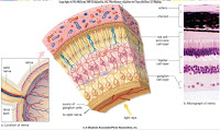
- Retina- located in posterior compartment (filled with vitreous humor). Contains rod cells and cone cells. Rods are sensitve to light but do not register color, and cones require bright light to recognize the different wavelengths (color). Fovea centralis is area of densely packed cells, and light is focused here when looking at an object. Sensory fibers from
 the retina form the optic nerve which in turn takes the impulses to the visual cortex.


The cornea is where focusing starts, and it continues through the lens and humors. Visual accomodation happens for close vision by the lens becoming more round to bring the object into focus.
Sense of Hearing
The two functions of the ear are hearing and balance. All of the sensory receptors of the ear contain hair cells with sereocilia sensitive to mechanical stimulation. These are mechanoreceptors. The ear is divided into three parts
- Outer- contains the pinna and auditory canal
- Middle- starts at the tympanic membrane and ends at the oval and round windows. The ossicles between the tympanic membrane and oval windows are: malleus, incus, stapes. The auditory tube allows the equalization of air pressure
- Inner- filled with fluid and broken down into semicircular canals, vestibule, and cochlea.
Hearing starts when sound waves enter the auditory canal and travel by vibrations to the tympanic membrane. The malleus takes this pressure and passes it using the incus to the stapes which hits the oval window membrane and passes the pressure to the fluid of the cochlea. In the cochlea the spiral organ has little hair cells and a tectorial membrane. Nerve impulses begin in the cochlear nerve and go to the brain. It is thought that the brain interprets tone based on the distribution of the stimulated hair cells
Sense of Equilibrium
The vestibular nerve works to achieve equilibrium and takes the nerve impulses to the cerebellum and brain stem. There are two types of mechanoreceptors involved in equilibrium
- Rotational- mechanoreceptors in semicircular canals detect angular and rotational movement. The three canals are arranged to each cover one dimension of space. The ampulla of each canal is enlarged and covered in little hair cells. As fluid in the canals moves over the ampulla, the hairs bend and the pattern of impulses being carried to the brain is changed.
- Gravitational- mechanoreceptors in the utricle and saccule detect verticle and horizontal movement. These are two membraneous sacs containing little hair cells in an otolithic membrane. When the head moves one way or another the otoliths are displaced and hair cells bent sending info to the brain. The brain uses the information it receives to maintain equilibrium by sending signals to the correct skelatal muscles to correct position
Works Cited
Mader, Sylvia. Human Biology 10th ed
Frolich powerpoint
Links to Pictures
1. http://www.virtualsciencefair.org/2004/visa4a0/public_html/brain6.gif
2. http://www.discoveryfund.org/images/Eye_Anatomy-Anat.jpg
3. http://www.biologyreference.com/images/biol_03_img0336.jpg
No comments:
Post a Comment