- Overview of Skeletal System
- Bone Growth, Remodeling and Repair
- Bones of the Axial Skeleton
- Bones of the Appendicular Skeleton
- Articulations
Muscular System
- Overview of the Muscular System
- Skeletal Muscle Fiber Contraction
- Whole Muscle Contraction
- Muscular Disorders
- Homeostasis
 re five functions of the skeletal system
- Supporting the body
- Protecting soft body parts
- Producing blood cells
- Storing minerals and fat
- Permit flexible body movement
A long bone is made up of a shaft (has medullary cavity with walls made of compact bone and filled with yellow bone marrow), epiphysis (spongey bone containing red bone marrow), and covered by a layer of periosteum. (contains blood and lympahtic vessels and nerves)
Compact bone is made of osteons which are tubular units. In the osteon, osteocytes lie in lacunae. Osteocytes exchange nutrients and wastes with the blood vessels in central canal.
Spongy bone is made up of many thin plates (trabeculae) separated by unequal spaces and designed for strength. Often contain red bone marrow.
Cartilage is not as strong as bone, but more flexible. This is due to the matrix being gel-like with collagen and elastic fibers. These cells are called chondrocytes. Cartilage has no nerves and no blood vessels. There are three types of cartilage
- Hyaline- firm and flexible
- Fibrocartilage- stronger and used in support
- Elastic- most flexible (ear flaps and epiglottis)
Fibrous connective tissue is made of rows of fibroblasts that are separated by bundles of collagen fibers. This is the tissue that ligaments and tendons are made of. Ligaments connect bone to bone and tendons connect muscles to bone at joints.
Bone Growth, Remodeling, and Repair
The skeleton starts to develop when the embryo is only 6 weeks old. Bones are made of living tissue and are able to grow throughout a lifetime by responding to stress (change shape, size and strength)
- Osteoblasts- are bone forming, encourage the deposition of calcium salts into the matrix
- Osteocytes- are mature bone cells that maintain structure
- Osteoclasts- are bone absorbing cells that break down bone and deposit calcium and phosphate into the blood.
Ossification is formation of bone. There are two different types of ossification
- Intramembranous- bones develop between sheets of fibrous connective tissue. These cells become osteoblasts and are found in ossification centers. They secrete the organic matrix and when calcium salts are added this results in calcification. Trabecule of spongy bone are formed and a periosteum develops around the outside. Compact bone is created on the outside. Example include skull bones and flat bones
- Endochondral- This is when cartilaginous models are replaced by real bones. Bone formation occurs from the center of the model to the end
- Cartilage model- chondrocytes put down hyaline cartilage which is the model
- Bone collar- Osteoblasts from the new periosteum secrete the matrix and then it undergoes calcification creating the bone collar to cover the diaphysis
- Primary ossification center- blood vessels carry osteoblasts to the interior to lay down spongy bone
- Medullary cavity and secondary ossification sites- spongy bones absorbed by osteoclasts and creates the medullary cavity
- Epiphyseal plate- band of cartilage that remains between primary ossification centers and each secondary center. Allows limbs to increase in length. There are four layers. First is the resting layer which remains cartilage, then the proliferating layer where chondrocytes produce new cartilage. Third is the degenerating zone where cartilage cells die and last is the ossification zone where bone is formed. When these plates close, the bone can no longer grow in length
Hormones are chemical messengers produced by one part of the body, but acting on another part. Vitamin D is converted into a hormone for the intestinal tract to help it absorb calcium. Growth hormone stimulates the epiphyseal plate and general bone growth. Thyroid hormone promotes metabolic activity in the cells.
Bone remodeling (bone renewal) keeps bones strong, while bone recycling allows regulation of calcium in the blood. The parathyroid hormone accelerates bone recycling and calcitonin is the hormone that acts the opposite way of parathyroid.
Bone repair is needed if a bone is fractured or broken. This repair can take several months and works in the following steps
- Hematoma- blood from the ruptured vessels forms a mass of clotted blood in between the broken bones
- Fibrocartilaginous callus- tissue repair starts and this callus fills in the space bewtween the broken part for up to three weeks
- Bony callus- Osteroblasts produce trabeculae and convert the fibrocartilaginous callus to a bony callus the joins the bones together for 3-4 months
- Remodeling- osteoblasts build new compact bone and osteoclasts absorb spongy bone which creates a new medullary cavity.
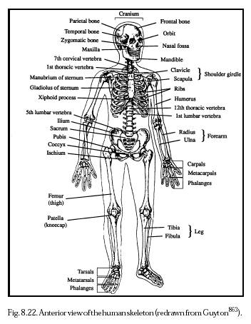
Bones of the Axial Skeleton
All of the skeletal bones are classified depending if they occur in the axial skeleton or appendicular skeleton. The axial skeleton is made of
Skull-
- Cranium ( protects the brain).
- Frontal bone (forms forehead),
- Parietal bone (sides)
- Occipital bone (base of the skull).
- Foramen magnum is the opening where the spinal cord passes and turns into the brain stem.
- Temperal bone (leads to middle ear),
- Sphenoid bone (extends across floor of cranium. all bones articulate with this one),
- Ethmoid bone (forms the nasal septum and orbits)
- Facial bones (mandible- only moveable bone, maxillae, zygomatic, nasal bones)
Hyoid Bone- only bone in the body that does not articulate with another bone. Attatched to temporal bones by muscle and ligaments and a membrane connects it to the larynx. Hyoid anchors the tongue and is place of attatchment for muscles used in swallowing
Vertebral Column- has 33 vertebrae that are named for their location in the vertebral column. First is the atlas (holds up the head). Second is the axis (rotation) then 12 thoracic, 5 lumbar, 5 sacrum, and 3-5 fused coccyx.
Rib Cage- also known as the thoracic cage is made of thoracic vertebrae, ribs, sternum, and cartilage. It protects the heart and moves with inspiration and expirtation.
Bones of the Appendicular Skeleton
These are bones in the pectoral and pelvic girdles and attatched limbs.
Pectoral girdle- there a right and left. Each one has a scapula, clavicle, glenoid cavity, rotator cuff, humerus, radius and ulna, carpal, metacarpals and phalanges.
Pelvic girdle- has two hip bones, pelvis, coxal bone ( ilium, ischium, pubis), pubic symphysis, femur, tibia, patella, fibula, tarsal, metatarsal and phalanges.
Articulations
Bones are joined together at joints (fibrous, cartilaginous, synovial) Most fibro us joints do not move. Cartilagenous joints are slightly moveable. Synovial joints move freely(flexion, extension, adduction, abduction, rotation, circumduction, inversion, eversion)
Muscular System
Overview of Muscular System
All muscles have the ability to contract causing movement. There are three ty pes of muscle
- Smooth muscle- spindle shaped cells with a single nucleus. Arranged in parrallel lines which form sheets. No striations. Located in walls of hollow internal organ and contraction is involuntary
- Cardiac muscle- form the heart wall. No nucleus, striated, tubular and branched which allows fibers to interlock. These fibers relax completely in between contractions which prevents fatigue. Involuntary contractions
- Skeletal muscle- fibers are tubular, have many nucleus' and are striated. They make up skeletal muscles and are voluntary.
Functions of the Skeletal muscles include
- Supporting the body- contraction opposes gravity and we remain upright

- Making bones move-muscle contraction results in movement
- Maintaining constant body temperature-contraction causes the breakdown of ATP which releases heat
- Assists movement in cardiovascular and lymphatic vessels- pressure of contraction keeps blood moving
- Protect internal organs and stabilize joints- pad bones, hold them together at joints and protect internal organs
Skeletal muscles are attatched to the skeleton. A muscle contains bundles of skeletal muscle fibers (fascicles). Each fiber is surrounded in connective tissue. Muscles are covered in fascia that goes past the muscle and becomes the tendon. These anchor a muscle to a bone. Usually each muscle works on the movement of one bone. These muscles work in pairs. Origin is on the stationary bone and insertion is on the bone that moves. Muscles can only pull, not push and this is why they work in pairs. A prime mover does most of the work with assistance from synergists and the antagonist is the muscle that works opposite the prime mover. One must relax while the oher is working.
The following terms help to characerize muscles
- Size- maximus, minimus, vastus, longus, brevis
- Shape- deltoid, trapezius, latissimus, terres
- Location- external, internal, frontalis,pectoralis, gluteaus,brachii, sub
- Direction of muscle fibers-rectus, orbicularis,transverse, oblique
- Attachment- sternocleidomastoid, brachioradialis
- Number of attachments-
- Action- extensor, adductor, flexor, masseter, levator
Skeletal Muscle Fiber Contraction
A muscle fiber is made up of a sarcolema(plasma membrane), sarcoplasm (cyt oplasm), sarcoplasmic reticulum (endoplasmic reticulum). Also the sarcolema creates T tubules which penetrate the cell to come into contact with expanded portions of the sarcoplasmic reticulum. These expanded sites are for calcium storage. The sarcoplasmic reticulum encases myofibrils and contains glycogen.
Myofibrils run the length of the muscle fiber. Sarcomeres are units of myofibrils and extends betweeen the Z lines. Contains myosin and actin. The I band has only actin and the  A band has myosin and actin overlapping. H zone has only myosin.
Thick filaments is hundreds of myosin molecules. Myosin is shaped like a golf club (the end is a cross bridge) Thin filaments is two intertwining strands fo actin. Sliding filaments let actin slide over myosin during contraction when the impulses travel down the T tubule into the sarcoplasmic reticulum where calcium is released. When sliding occurs, sarcomeres shorten and the myosin filaments break down ATP.
Muscle fibers contract when stimulated by motor neurons with there axons i n nerves. Axon terminals have synaptic vesicles filled with acetylcholine. When the nerve impulses reach the axon terminal, synaptic vessels release ACh into the cleft. When this is released it diffuses across the cleft and binds to sarcolemma receptors. Then impulses spread through T tubules to reticulum and Calcium is released causing sarcomere contraction. Calcium binds with troponin, and myosin binding sites are exposed allowing the binding to actin.
Whole Muscle Contraction
A nerve fiber with all the muscle fibers in innervates is a motor unit. This uni t follows the all or none law. A muscle twitch is when a motor unit is stimulated by infrequent electrical impulses. Tetanus is maximum sustained contraction. This will continue until muscle fatigue occurs.
Muscles use different fuel sources for energy and different ways to produce ATP during contraction. Glycogen and fat are stored in muscle while blood glucose and plasma fatty acids come from the blood. Muscles acquire ATP by the creatine phosphate pathway, fermentation, can cellular respiration
Fast twitch fibers metabolize anaerobically and are designed for strength due to the fact their motor units contain many fibers and provide explosions of energy. They develop maximum tension more rapidly, but are more susceptible to accumulation of lactate
Slow twitch fibers have more endurance and produce energy aerobically causing them to tire when their fuel supply is gone. Contain myoglobin and lots of mitochondria. Surounded by capillary beds and draw more blood and oxygen.
Muscular Disorders
Common disorders
- Spasms- sudden, involuntary muscular contraction
- Cramps- strong, painful spasms
- Strain- stretching or tearing a muscle
- Sprain- twisting of a joint, includes damage to ligaments, tendons, blood vessels, nerves
- Tendinitis- gliding motion of tendon impaired
- Bursitis- inflamation of bursa
Serious conditions
- Myalgia-achy muscles due to overuse or over stretching
- Muscular dystrophy- group of disorders characterized by progressive degeneration
- Myasthenia gravis- autoimmune disease characterized by weakness. Muscle contraction is impaired when the immune system accidentally produces antibodies that destroy ACh
- Amyotrophis lateral sclereosis- gradual loss of ability to walk, talk, chew, and swallow
Homeostasis
Muscle movement allows us to respond to environmental changes, allow us to eat, to digest that which we eat to provide cells with nutrients, breathe, circulate blood and lymph. Also the skeletal and muscle systems protect internal organs allowing them to carry out their part of homeostasis. The skelaton is largely involved in calcium homeostasis. Also blood cells are produced in bones in the red bone marrow where white blood cells are also produced. Muscles help maintain body temperature (shivering, goosebumps)
Works Cited
Mader, Sylvia. Human Biology 10th ed
Frolich Powerpoint
Links for Pictures
1. http://www.web-books.com/eLibrary/Medicine/Physiology/Skeletal/long_bone.jpg
2. http://www.nanomedicine.com/NMI/Figures/8.22.jpg
3. http://www.pinkmonkey.com/studyguides/subjects/biology-edited/chap20/fig20_2.gif
4. http://picinfor.googlepages.com/02620Types20of20joints20found20in20the20human20body.jpg/02620Types20of20joints20found20in20the20human20body-full.jpg
5. http://virtualastronaut.jsc.nasa.gov/textonly/act21/images/musclemap.png
6. http://www.agen.ufl.edu/~chyn/age2062/lect/lect_19/146.gif
7. http://www.mhhe.com/biosci/esp/2001_gbio/folder_structure/an/m5/s5/assets/images/anm5s5_1.jpg
8. http://www.rogers.k12.ar.us/users/ehutches/musaction6.jpg
9. http://fig.cox.miami.edu/~cmallery/150/neuro/neuromuscular-sml.jpg
10. http://www.edcenter.sdsu.edu/cso/paper/image005.jpg
No comments:
Post a Comment